
This article is written for dentists and should not be read as patient information or a treatment recommendation. It is a literature-based clinical guide by Dr. Ferit Çiftçi and has not been peer reviewed. References and a methods note are at the end.
CLINICAL GUIDE · IMPLANTS AND PROSTHODONTICS
Accuracy in implant impressions: tray technique, splinting and intraoral scanning
CLINICAL SUMMARY
In a 2014 review of conventional impressions, the splinted technique was more accurate in both partially and completely edentulous patients, and the open tray was ahead in the edentulous arch. On that question half of the laboratory studies found no difference, and another review focused on the edentulous arch could make no clear recommendation.1, 14 For two-unit restorations on two non-parallel implants, a limited number of laboratory studies found splinting the impression copings unnecessary.2 Intraoral scanning gives accuracy comparable to a conventional impression for single units and short spans, and has limitations in long spans and in extended partial or complete edentulism.3 In a complete arch with more than four implants, conventional impressions came out slightly ahead.4 Most of the evidence comes from laboratory studies.1, 5 A verification jig used selectively before the definitive prosthesis can reduce the likelihood of misfit.6
Where does misfit start?
Misfit in a multi-implant framework comes from the sum of small errors made during impression-taking, cast fabrication and milling.6 According to a 1995 review, impression making, the master cast, the resin verification jig, waxing, investing, casting, veneer addition and finishing can all add to the final distortion of the prosthesis. In the author's view, a totally passive fit is probably unachievable; the task is to define a level of distortion and associated stress that is acceptable and gives the prosthesis adequate clinical longevity.7 Scanning has the same problem: in complete-arch intraoral scans the recorded position of the scan bodies can deviate markedly from their actual position, and the definitive prosthesis then does not fit passively.8
There is no agreed clinical threshold for acceptable misfit. A systematic review from the consensus project of the European Prosthodontic Association (EPA) states that this threshold, and objective criteria for assessing linear and angular deviation, still need to be verified in clinical studies.9 In this article, "more accurate" means that the measured deviation was smaller. There are not enough clinical data to say how much that difference matters in the mouth.5
Conventional impression or scan?
The sources separate the methods mainly by three facts: how extensive the edentulism is, how many implants there are, and the angle between them. According to the German S2k guideline on intraoral scanning, scanning gives accuracy comparable to conventional methods for tooth- or implant-supported single units and short-span restorations. It has limitations in long-span prostheses of more than four units and in extended partially or completely edentulous cases. The guideline also states that clinical performance depends on the indication, the workflow and the operator's experience.3
In a review of seven clinical studies in 151 partially dentate patients, four studies entered the meta-analysis of deviation and three that of angular displacement: deviation was significantly smaller with digital impressions, while angular displacement did not differ. Heterogeneity between studies was high, and the authors advise caution in long-span cases and with angulated implants.10 The 2021 EAO consensus report states that patient comfort is greater with optical impressions for implant-supported single crowns and short-span bridges.11
In the edentulous arch the picture is less clear. A 2026 review of 37 studies found, in a meta-analysis of 25 of them, no overall difference between digital and conventional impressions. There was no difference with four implants or fewer; with more than four, conventional impressions were slightly ahead. Photogrammetry was slightly more accurate than conventional impressions, and intraoral scanners did not differ from them. Certainty of evidence was moderate, and there were signs of publication bias.4 The 2018 ITI consensus report states that with an intraoral scanner in edentulous cases, the result depends on the protocol followed.12 In a randomised pilot trial of 22 patients receiving complete-arch implant prostheses, framework passive fit was similar with both methods, and both patients and operators found scanning more comfortable. The trial enrolled cases with predefined limits on implant number, spacing and angulation.13
-
How extensive is the edentulism?
- PARTIAL
Long span or angulated implants?
- NO
- YES
- EDENTULOUS ARCH
More than four implants?
- NO
- YES
- PARTIAL
Tray, splint and material
In a 2014 systematic review of 76 studies (4 clinical, 72 laboratory), most laboratory studies and all three clinical studies in edentulous patients found the splinted technique more accurate (15 studies in favour of splinting, 1 in favour of non-splinting, 9 with no difference). The open tray was more accurate than the closed tray in 10 studies, 1 study favoured the closed tray, and 10 found no difference, so half of the studies on this question showed no difference. In partial edentulism the splinted technique was also ahead (8 studies in favour of splinting, 2 in favour of non-splinting, 3 with no difference). The open tray led in most laboratory studies there, but the only clinical study in that group found no difference. Polyether and polyvinyl siloxane did not differ in accuracy; implant angulation affects accuracy.1
Another review published the same year and focused on the edentulous arch (34 studies, one clinical) found the evidence on tray and splinting technique conflicting and made no clear recommendation. Of 24 studies on the splinting effect, 10 found no difference and 7 found splinting superior.14 In the edentulous arch, a splinted open tray is the choice that follows the trend of the evidence; it has not been shown to make a difference in every case.
The review of two-unit restorations on two non-parallel implants included five laboratory studies, four of them in the meta-analysis. According to the results section of the abstract, splinted copings gave statistically smaller marginal gaps and made no difference to framework strain; the conclusions section, however, states that marginal gaps were significantly larger with splinted copings. The abstract contradicts itself on this point, and the full text was not read for this article. All studies used internal-connection implants and implant-level impressions, with angulation between 8 and 30 degrees. The authors judged the clinical significance low and, based on the limited number of studies, considered splinting unnecessary in this situation.2
In most situations the impression material does not change accuracy. In a 2025 review on implant-supported fixed complete dentures, 27 laboratory studies entered the qualitative and 12 the quantitative analysis; polyvinyl siloxane and polyether gave similar accuracy. When implants were angled more than 15 degrees, a difference in favour of polyether was found; for the closed tray this result rests on only two studies. The finding comes from the laboratory and from complete-arch fixed prostheses only.15
Scan bodies and complete-arch scanning
According to a 2024 review of 16 studies, scan accuracy depends on the position of the scan body in the arch and on its design, shape, material, colour and manufacturing system. The authors list scanner type, scanning technique, scan aids, inter-implant distance, scan span and the number of implants as further factors that warrant evaluation. For single implants and short spans, scanning gives accuracy similar to traditional impressions; there are not enough in vivo data for clinical recommendations.16 The 20 studies reviewed separately on scan body design could not reach a concrete conclusion on which design is most accurate.17
Scan body seating is sensitive to how the scan body is tightened. On a single-implant laboratory model, the vertical deviation of the scan body varied with operator level and with tightening torque. The largest mean deviation, 13.5 µm, was measured at 35 Ncm, and none of the deviations exceeded a previously described occlusal threshold. The study compared hand tightening with 15 Ncm; the abstract does not say which was better.18 No torque value follows from the study; what follows is that the tightening method and the operator changed the vertical position. Tighten every scan body by the method in the system's instructions, the same way each time.
Seating the scan body on the implant
-
CORRECT
The scan body sits on the implant platform with no gap, and its axis follows the implant. No error from seating is added to the record.
-
INCORRECT
There is a gap at the interface and the scan body is tilted. The scanner records this position, and the error is carried into the framework.
In the complete arch, additional techniques can improve accuracy. In a review of 17 techniques, the additional techniques were more accurate than the standard scanning protocol, and in most studies the common element was a splinting device connecting the scan bodies. In return they need extra devices, more scans and more time.8 A review comparing splinted and non-splinted scan bodies also reports that splinting improves accuracy, especially in complete-arch cases, eases the stitching of images and gives more stable reference points. Light-cured resin, pattern resin, dental floss and custom-made splints were used; some studies found no improvement or even a negative effect.20 For scanning edentulous arches, the factors listed are scanner type, scanning technique, environmental conditions, the angle and distance between implants and the scan body material. Creating physical paths that join the scan bodies can improve the transfer of implant positions, and PEEK scan bodies were more accurate.21
Results vary widely between studies. In a meta-analysis of 100 studies on non-splinted complete-arch scans, linear discrepancy ranged from 22 to 1050 µm (mean 117 µm), with significant publication bias.22 A review of 12 studies on commercially available non-calibrated splinting methods found significant differences between methods, all within the 150 µm threshold that the authors consider clinically acceptable; the evidence is limited.23
The effect of angulation on scanning is debated. A review of eight laboratory studies advises caution with scanning when the angle between implants exceeds 15 degrees.24 In the studies that measured RMS (root mean square deviation) in a 49-study meta-analysis, however, scanning was more accurate than conventional impressions for non-parallel implants, and conventional impressions were slightly more accurate for parallel implants.25
Verification before the framework
An implant verification jig has been described as a quality-control device in fixed implant prosthodontics. According to a 2025 narrative review of 52 publications (12 laboratory and 8 clinical studies), jigs can be used as an adjunct to conventional impressions and to intraoral scans with vertical scan bodies. PMMA, UDMA, light-polymerising composite resins and Type III/IV dental stone have been used; digital workflows describe scanning the jig or a verification cast, and CAD-CAM jigs. Photogrammetry gives more accurate data than intraoral scanners, and systems that pair horizontal scan bodies with AI-based recognition also report more accurate data, although published studies on them remain few; a conventional jig may not be needed with those methods. The review considers verification prudent particularly with long spans between implants and no fixed keratinised mucosa, and notes that many prostheses made from digital scans are monolithic full-contour restorations, which leaves no opportunity for a traditional framework try-in. The authors state that selective use before the definitive prosthesis can reduce the likelihood of misfit.6 The jig itself can add distortion: the 1995 review lists the resin verification jig among the stages that can add to it.7
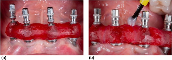
At try-in, assess fit with several separate checks. In the randomised pilot trial on complete-arch implant prostheses, passive fit was scored with five components: framework lift-off, the single-screw test, the full-screw test, smoothness of screw insertion and the radiographic gap.13 In the single-screw test (Sheffield test), the screw on one terminal implant is tightened and the other implants are checked for a gap or lift-off (Figure 3).6 The same five components can serve as a checking sequence at the chair. There is a limit: soft-tissue interference, saliva, screw preload and limited access with subgingival and internal connections can mask misfit.6
Step-by-step protocol
Choose the method for the case
Note the extent of edentulism, the number of implants and the angle between them. For single units and short spans, scans and impressions give comparable accuracy.3 If you take a conventional impression in the edentulous arch, the trend of the evidence favours a splinted open tray, although results conflict.1, 14
Account for angulation in advance
For complete-arch fixed prostheses, polyether was ahead in the laboratory beyond 15 degrees (material).15 For scanning, the evidence on angulation is conflicting.24, 25
Seat the components and check seating
Tighten scan bodies and impression copings by the method in the system's instructions, the same way each time.18 If the seating of an impression coping is in doubt, take the radiograph with the beam perpendicular to the long axis of the implant.19
Splint according to the case
In the 2014 review, splinting was ahead in partial and complete edentulism.1 For a two-unit restoration on two non-parallel implants it was found unnecessary in the laboratory.2
Tie complete-arch scans to a protocol
The result depends on the protocol followed.12 If you will use an aid that connects the scan bodies, plan the extra devices and time in advance (scanning).8, 20
Verify before the framework
Check implant positions with a verification jig or a verification cast.6 At try-in, record all five components: lift-off, the single-screw test, the full-screw test, smooth screw insertion and the radiographic gap.13
Errors and corrections
| Error | Correction |
|---|---|
| Taking a non-splinted closed-tray impression in the edentulous arch. | The trend of the evidence favours the splinted technique and the open tray (tray and splint).1 |
| Applying the "splinting is unnecessary" finding for two-unit restorations to every case. | The finding rests on laboratory measurements of two-unit restorations on non-parallel, internal-connection implants with implant-level impressions.2 |
| Tightening scan bodies differently each time, or having different people tighten them. | The tightening method and the operator changed the vertical position; tighten every scan body by the method in the instructions, the same way each time.18 |
| Planning a complete-arch scan as if it were a short-span scan. | Scanning has limitations in long spans and in the edentulous arch;3 decide the protocol and, if needed, an aid connecting the scan bodies beforehand (scanning). |
| Noticing angulation only after the impression is taken. | Angulation affects impression accuracy;1 note the angle before the impression and choose the material accordingly (material).15 |
| Moving to the definitive framework without verification. | With a long span and no fixed keratinised mucosa, do not skip verification; at try-in, check all five components (verification).6, 13 |
Level of evidence
What is known
Polyether and polyvinyl siloxane give similar results in most situations.1, 15 Intraoral scanning gives accuracy close to conventional impressions for single units and short spans and increases patient comfort.3, 11 In the complete arch, digital and conventional impressions are generally similar; with more than four implants conventional impressions are slightly ahead, while photogrammetry was slightly more accurate than conventional impressions.4 In the review from the EPA consensus project, intraoral scanning and photogrammetry showed comparable accuracy for recording implant positions in the edentulous arch.9
What is not known
Most of the evidence is from the laboratory: 72 of the 76 studies in the 2014 review and 75 of the 79 studies in the 2018 review prepared for the ITI were in vitro. The second review states that in vivo data are insufficient for clinical recommendations.1, 5 Results on tray and splinting technique conflict: in one 2014 review splinting was ahead in partial and complete edentulism, but on the open-tray question in the edentulous arch half of the studies found no difference; in the other, 10 of 24 studies on the splinting effect found no difference and the author made no clear recommendation.1, 14 The abstract of the meta-analysis on two-unit restorations contradicts itself.2 An acceptable misfit threshold and objective assessment criteria have not been verified in clinical studies.9 The most accurate scan body design has not been identified.17 Complete-arch scanning results vary widely between studies, with signs of publication bias.22, 4 How verification jigs should be used has not been standardised, and their long-term clinical outcomes have not been evaluated.6 The randomised pilot trial found in this search is limited to 22 patients and six months of follow-up.13
At the chair
References
- Papaspyridakos P, Chen CJ, Gallucci GO, Doukoudakis A, Weber HP, Chronopoulos V. Accuracy of implant impressions for partially and completely edentulous patients: a systematic review. Int J Oral Maxillofac Implants. 2014;29(4):836-45. PMID: 25032763 · DOI: 10.11607/jomi.3625
- Ma Y, Al-Radhi RYZ, Jiang L, Yu H. Does Splinting the Direct Copings Increase the Impression Accuracy of Two-Unit Nonparallel Implant Restorations? A Systematic Review and Meta-analysis. Int J Oral Maxillofac Implants. 2022;37(4):653-659. PMID: 35904821 · DOI: 10.11607/jomi.9577
- Ruhstorfer M, Graf T, Baresel I, Flügge T, Reich S, Reiss B, et al. Results of the 'German S2k guideline on intraoral scan in dental medicine' - Part II: Clinical Applications. Int J Comput Dent. 2026. Epub ahead of print. PMID: 42820673 · DOI: 10.3290/j.ijcd.b7114807 · Guideline record: DGCZ/DGZMK. Intraoralscan in der Zahnmedizin. S2k-Leitlinie, Langversion 1.0, 2025. AWMF register no. 083-049: register.awmf.org
- Alfaraj A, Alqudaihi F, Khurshid Z, Qadiri O, Lin WS. Comparative analyses of accuracy between digital and conventional impressions for complete-arch implant-supported fixed dental prostheses-A systematic review and meta-analysis. J Prosthodont. 2026;35(3):252-274. PMID: 40660725 · DOI: 10.1111/jopr.14094
- Flügge T, van der Meer WJ, Gonzalez BG, Vach K, Wismeijer D, Wang P. The accuracy of different dental impression techniques for implant-supported dental prostheses: A systematic review and meta-analysis. Clin Oral Implants Res. 2018;29 Suppl 16:374-392. PMID: 30328182 · DOI: 10.1111/clr.13273
- Wong AWH, Nedelcu R, Hamilton A. Clinical Recommendations for Implant Verification Jigs in Analogue and Digital Workflows. A Narrative Review. Aust Dent J. 2025;70 Suppl 1:S35-S49. PMID: 41288068 · DOI: 10.1111/adj.70025
- Tan KB. The clinical significance of distortion in implant prosthodontics: is there such a thing as passive fit? Ann Acad Med Singap. 1995;24(1):138-57. PMID: 7605083 · DOI: not listed in PubMed
- Paratelli A, Vania S, Gómez-Polo C, Ortega R, Revilla-León M, Gómez-Polo M. Techniques to improve the accuracy of complete arch implant intraoral digital scans: A systematic review. J Prosthet Dent. 2023;129(6):844-854. PMID: 34756427 · DOI: 10.1016/j.prosdent.2021.08.018
- Rutkūnas V, Gedrimienė A, Mischitz I, Mijiritsky E, Huber S. EPA Consensus Project Paper: Accuracy of Photogrammetry Devices, Intraoral Scanners, and Conventional Techniques for the Full-Arch Implant Impressions: A Systematic Review. Eur J Prosthodont Restor Dent. 2023. Epub ahead of print. PMID: 37314199 · DOI: 10.1922/EJPRD_2481Rutkunas12
- Park JS, Alshehri YFA, Kruger E, Villata L. Accuracy of digital versus conventional implant impressions in partially dentate patients: A systematic review and meta-analysis. J Dent. 2025;160:105918. PMID: 40553823 · DOI: 10.1016/j.jdent.2025.105918
- Jokstad A, Pjetursson BE, Mühlemann S, Wismeijer D, Wolfart S, Fehmer V, et al. Fabrication, workflow and delivery of reconstruction: Summary and consensus statements of group 4. The 6th EAO Consensus Conference 2021. Clin Oral Implants Res. 2021;32 Suppl 21:336-341. PMID: 34145922 · DOI: 10.1111/clr.13797
- Wismeijer D, Joda T, Flügge T, Fokas G, Tahmaseb A, Bechelli D, et al. Group 5 ITI Consensus Report: Digital technologies. Clin Oral Implants Res. 2018;29 Suppl 16:436-442. PMID: 30328201 · DOI: 10.1111/clr.13309
- Liu M, Shi JY, Fu XJ, Liu BL, Qiao SC, Lai HC. Comparison of short-term clinical outcomes and patient satisfaction between intraoral scanning and conventional impressions for complete-arch implant prostheses: a pilot RCT. J Dent. 2026;174:106887. PMID: 42425293 · DOI: 10.1016/j.jdent.2026.106887
- Baig MR. Accuracy of impressions of multiple implants in the edentulous arch: a systematic review. Int J Oral Maxillofac Implants. 2014;29(4):869-80. PMID: 25032767 · DOI: 10.11607/jomi.3233
- Guo YQ, Ma Y, Cai SN, Yu H. Optimal impression materials for implant-supported fixed complete dentures: A systematic review and meta-analysis. J Prosthet Dent. 2025;133(5):1172-1182. PMID: 37599185 · DOI: 10.1016/j.prosdent.2023.06.024
- Gehrke P, Rashidpour M, Sader R, Weigl P. A systematic review of factors impacting intraoral scanning accuracy in implant dentistry with emphasis on scan bodies. Int J Implant Dent. 2024;10(1):20. PMID: 38691258 · DOI: 10.1186/s40729-024-00543-0
- Gómez-Polo M, Donmez MB, Çakmak G, Yilmaz B, Revilla-León M. Influence of implant scan body design (height, diameter, geometry, material, and retention system) on intraoral scanning accuracy: A systematic review. J Prosthodont. 2023;32(S2):165-180. PMID: 37771200 · DOI: 10.1111/jopr.13774
- Shi X, Liu X, Liu S, Wang M, Liu F. Vertical Deviation Caused by Tightening Torque on Implant Scan Body: An In Vitro Study. Int J Prosthodont. 2022;35(5):653-659. PMID: 35649276 · DOI: 10.11607/ijp.7493
- Sharkey S, Kelly A, Houston F, O'Sullivan M, Quinn F, O'Connell B. A radiographic analysis of implant component misfit. Int J Oral Maxillofac Implants. 2011;26(4):807-15. PMID: 21841991 · DOI: not listed in PubMed
- Shetty PS, Gangurde AP, Chauhan MR, Jaiswal NV, Salian PR, Singh V. Accuracy of the digital implant impression with splinted and non-splinted intraoral scan bodies: A systematic review. J Indian Prosthodont Soc. 2025;25(1):3-12. PMID: 40654117 · DOI: 10.4103/jips.jips_261_24
- Carneiro Pereira AL, Souza Curinga MR, Melo Segundo HV, da Fonte Porto Carreiro A. Factors that influence the accuracy of intraoral scanning of total edentulous arches rehabilitated with multiple implants: A systematic review. J Prosthet Dent. 2023;129(6):855-862. PMID: 34656307 · DOI: 10.1016/j.prosdent.2021.09.001
- Revilla-León M, Barmak AB, Prasad S. Accuracy of complete arch implant scans using nonsplinting techniques: A systematic review and meta-analysis. Report of the Committee on Research in Fixed Prosthodontics of the American Academy of Fixed Prosthodon. J Prosthet Dent. 2026;136(1):e102-e138. PMID: 41850947 · DOI: 10.1016/j.prosdent.2026.02.034
- Revilla-León M, Ghunaim D, Barmak AB, Afshari FS, Fang Q, Prasad S. A systematic review of the accuracy of complete arch implant scans recorded by using noncalibrated splinting or implant scan body techniques. Report of the Committee on Research in Fixed Prosthodontics of the American Academy of Fixed Prosthodontics. J Prosthet Dent. 2026;135(6):1058-1071. PMID: 41345005 · DOI: 10.1016/j.prosdent.2025.11.018
- Carneiro Pereira AL, Medeiros VR, da Fonte Porto Carreiro A. Influence of implant position on the accuracy of intraoral scanning in fully edentulous arches: A systematic review. J Prosthet Dent. 2021;126(6):749-755. PMID: 33268069 · DOI: 10.1016/j.prosdent.2020.09.008
- Cai ZZ, Li X, Wu XY, Lai HC, Shi JY. Does intra-oral scan improve the impression accuracy of full-arch implant-supported prostheses: A systematic review and meta-analysis. Clin Implant Dent Relat Res. 2024;26(5):847-861. PMID: 38517307 · DOI: 10.1111/cid.13321
Methods note
Sources were searched in PubMed (NCBI E-utilities) on 5 November 2026. The search covered open and closed trays, splinting of impression copings, impression materials, implant angulation, digital and conventional implant impressions, scan body design and seating, complete-arch scanning techniques, photogrammetry and verification jigs. Systematic reviews, meta-analyses, consensus reports and guidelines were prioritised; two laboratory studies on scan body seating and radiographic checking, and one randomised pilot trial on assessing passive fit at try-in, were added. The German S2k guideline (AWMF register no. 083-049) is cited from its published journal summary; the AWMF register page could not be opened because of a bot check, and the register number was taken from a professional journal's announcement of the guideline. For each source, the authors, title, journal, year and DOI were checked against the PubMed record. Findings are taken from the published abstracts; the full text of the verification jig review (6) was read in PMC, and the limits on verification and try-in come from it. The caption and CC BY 4.0 licence of Figure 3 were verified in the same PMC record. The other open-access full texts (16, 20) were not read, and the contradiction in the abstract of reference 2 was not resolved from the full text. No textbook was used. No product or brand names are given in the text. This is not a systematic review; it was prepared by a single reader.
This text is not patient information. Treatment decisions are made after an examination and for each person individually.
Phone: +90 538 073 12 11